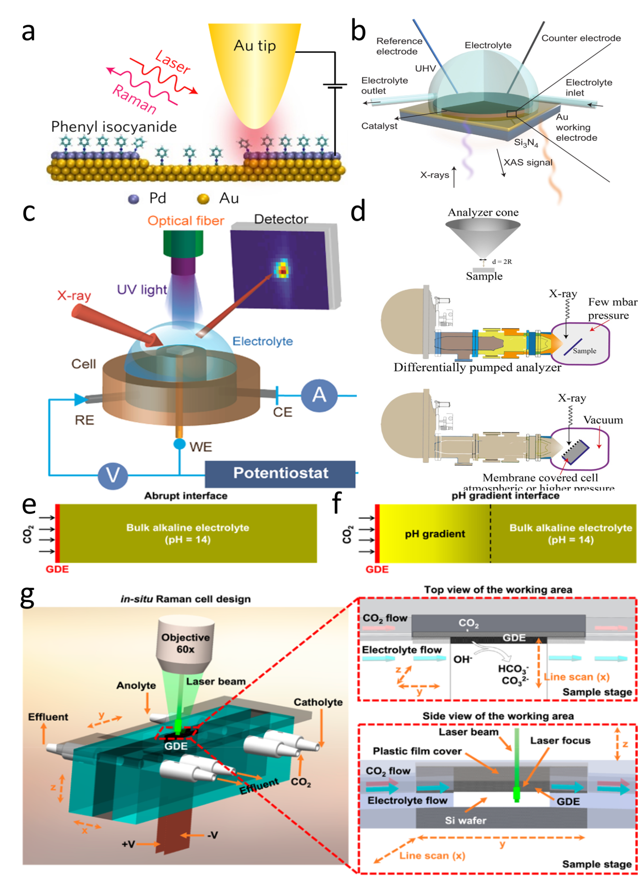
Figure 9. (a) Schematic of an STM-based time-dependent tip-enhanced Raman spectroscopy (TERS).[140] Copyright 2016, Nature Publishing Group. (b) Schematic illustration of the in situ XAS liquid cell.[142] Copyright 2018, Nature Publishing Group. (c) Schematic of the experimental setup for operando X-ray reflectivity of SrTiO3.[141] Copyright 2016, American Chemical Society. (d) Schematic illustration of the in situ XPS experimental set-up.[143] Copyright 2018, Wiley-VCH Verlag GmbH & Co. Designed flow cell for performing in situ Raman measurements to distinguish between (e) an abrupt interface and (f) a gradient interface. (g) The cell design with both top and side views of the cathode area.[144] Copyright 2020, American Chemical Society.
Other figure/table from this article